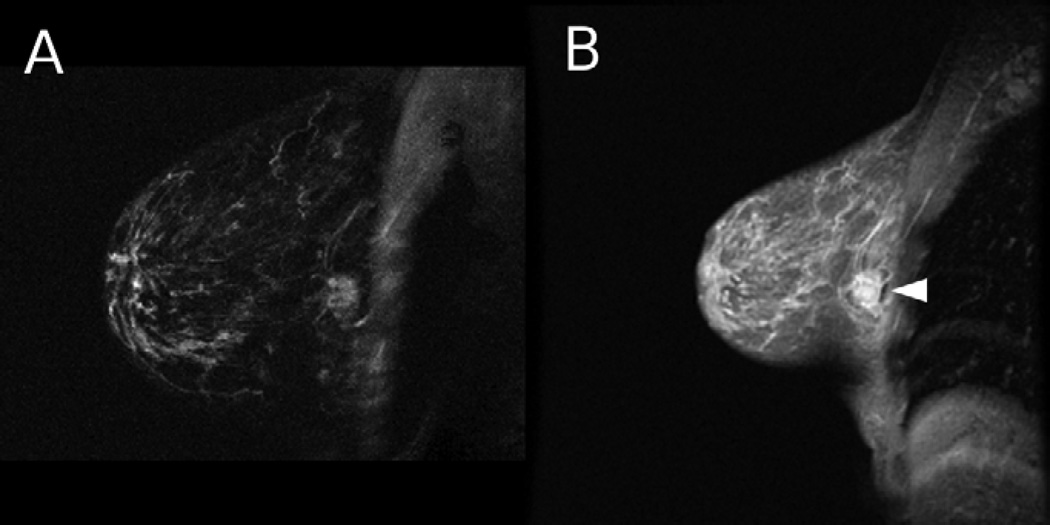
Figure 3.
A 44 year old woman with a cancerous lesion and lymph node invasion (not shown) was imaged using HiSS and conventional imaging. a) HiSS water peak height (TR/TE = 250/96 ms, in-plane resolution 0.63 mm) and b) conventional T1-weighted (TR/TE = 175/4.2 ms, in-plane resolution 1 mm) sagittal images are shown. The lesion is indicated with an arrow. Fat suppression is superior in the HiSS image.

