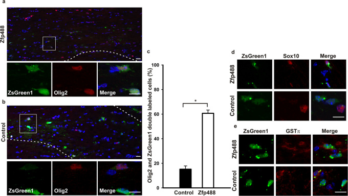
Figure 3. Mature oligodendrocytes derived from Zfp488 overexpressing SVZ progenitor cells survive after complete recovery from cuprizone-induced demyelination.
Mice on a 0.2% cuprizone diet for 2 weeks were injected with either control or Zfp488 expressing retrovirus into the SVZ. After cuprizone treatment for 5 weeks, mice were allowed to recover and remyelinate for 6 weeks on normal chow. After 5 weeks in the cuprizone diet, brain sections were evaluated for the differentiation status of the virus-transduced SVZ progenitor cell population. (a) Representative image of the Zfp488 virus transduced SVZ-derived cells in the white matter of the corpus callosum. Higher magnification panel show that cells transduced with the Zfp488 retrovirus differentiated into oligodendrocytes and were positive for Olig2. (b) Representative image of the control (ZsGreen1) virus transduced cells in the white matter of the corpus callosum. Higher magnification panel show that cells transduced with the control retrovirus did not differentiate into oligodendrocytes and were negative for Olig2. (c) Percentage of virus-transduced SVZ-derived cells that express Olig2. The number of ZsGreen1 positive cells that were positive for Olig2 was significantly higher in mice expressing Zfp488 (n=5) compared to controls (n=4, p<0.001). (d) SVZ-derived cells in the corpus callosum after 5 weeks of cuprizone-induced demyelination followed by 6 weeks of recovery. Representative images showed that Zfp488 expressing cells were also positive for the oligodendroglial marker Sox10 compared to control cells that were negative for Sox10. (e) SVZ-derived cells in the corpus callosum after 5 weeks of cuprizone-induced demyelination followed by 6 weeks of recovery. Representative images showed that Zfp488-expressing cells were also positive for the mature oligodendrocyte marker GSTπ compared to control cells that were negative for GSTπ. Scale bars, 100 μm and 50 μm for the lower and higher magnification images, respectively.

