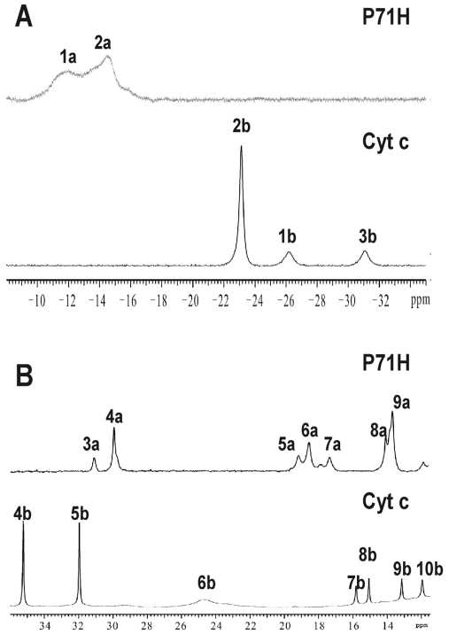
Figure 3.
(A) The high-field shifted region of 1D 1H NMR spectra of the native cyt c (down) and its P71H variant (upper) in oxidized state. (B) The down-field shifted region of 1D 1H NMR spectra of the native cyt c (down) and its P71H variant (upper) in oxidized state. The peaks in 1D 1H-NMR spectrum were assigned as (1a) His18 Hε1, (2a) His71 Hε1, (3a) His71 Hδ2, (4a) heme 8-CH3, (5a) His18 Hδ2, (6a) heme 3-CH3, (7a) His71 Hδ1, (8a) His18 Hδ1 and (9a) heme 5-CH3, respectively. The peaks in 1D 1H-NMR spectrum of native cyt c were assigned as (1b) His18 Hε1, (2b) Met80 ε-CH3, (3b) Met80 Hγ, (4b) heme 8-CH3, (5b) heme 3-CH3, (6b) His18 Hδ2, (7b) heme 7-Hα2, (8b) His18 Hβ2, (9b) heme 7-Hα1 and (10b) His18 Hδ1, respectively.

