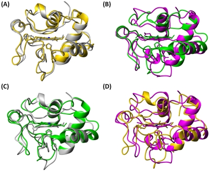
Figure 6.
Upon overlaying Cα atoms in second structural region and heme backbone atoms, the conformational comparison: (A) between the oxidized (grey, pdb code 1YIC) and the reduced (gold, pdb code 1YFC) native cyt c; (B) between the oxidized (green) and the reduced (magenta) P71H mutant; (C) between the oxidized native cyt c (grey, pdb code 1YIC) and P71H (green); (D) between the reduced native cyt c (gold, pdb code 1YFC) and the reduced P71H mutant (magenta).

