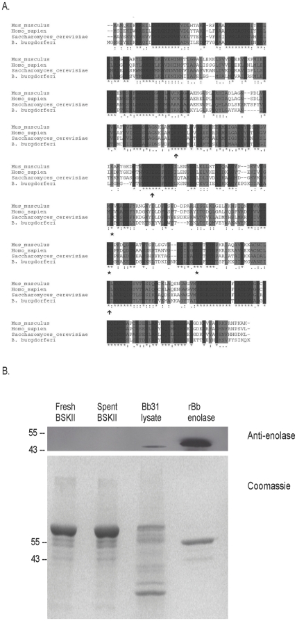
Figure 1. Conservation of enolase.
(A) Alignment of enolases from mouse, human, yeast and B. burgdorferi. Dark shaded residues indicate identity; light gray residues indicate similarity. Arrows indicate the 3 conserved residues that make up the enzymatic active site; stars indicate the three conserved residues required for binding the cofactor manganese. (B) A rabbit polyclonal antiserum directed against human enolase recognizes enolase from B. burgdorferi. Top panel: Western blot; Bottom panel: Coomassie-stained SDS-PAGE gel. Molecular weight markers are indicated on the left. Lane 1: fresh BSKII medium; Lane 2: spent BSKII medium from late-log phase cultures of B. burgdorferi; Lane 3: B. burgdorferi cell lysate; Lane 4: recombinant enolase from B. burgdorferi.

