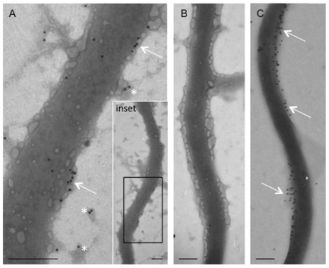
Figure 3. Immunoelectron microscopic analysis of B. burgdorferi enolase.
A) Anti-enolase was localized intermittently across the outer surface of B. burgdorferi (arrows). Gold particles were also observed on membrane blebs in proximity to the spirochete (asterisks). The boxed area in the inset indicates the region demonstrated in image (A). B) Omission of the primary antibody resulted in a complete loss of immunoreactivity. C) Anti-OspC immunolabeling demonstrated moderately heavy labeling on the outer surface of the spirochete (Arrows). Magnification bar = 0.2 µm.

