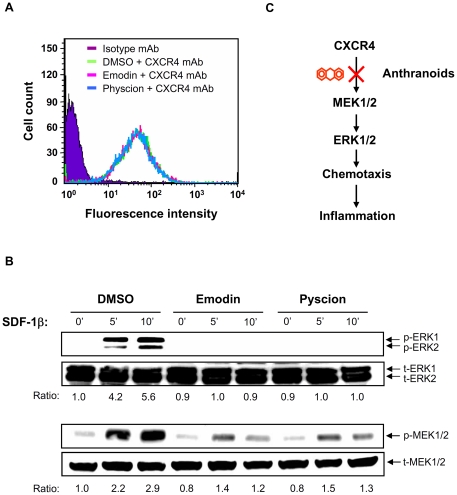
Figure 4. Effect of emodin and physcion on CXCR4 signaling.
A. Jurkat cells were incubated with emodin (1 µg/ml), physcion (1 µg/ml) or DMSO vehicle for 1 h at 37°C. After extensive washing, the cells were stained with anti-CXCR4 antibody or isotype antibody (control), followed by FITC-conjugated secondary antibody. The surface expression level of CXCR4 is shown as logarithmic fluorescence intensity. Data are representative examples of three independent experiments. B. The cells, with the same pre-treatment as Figure 4A, were stimulated with SDF-1 (100 ng/ml) from 0 to 15 min. Total lysates were analyzed by Western blot with antibody against ERK1/2 (t-ERK1/2) and their phosphorylated proteins (p-ERK1/2) (upper panel). After stripping, the same membrane was re-blotted with the antibody against MEK (t-MEK1/2) and their phosphorylated proteins (p-MEK1/2) (lower panel). The ratio was obtained by normalizing the signal of the phosphorylated protein to that of the total protein. C. Working model showing that the anthranoids, emodin and physcion, inhibit CXCR4-implicated chemotaxis and, in turn, inflammation.

