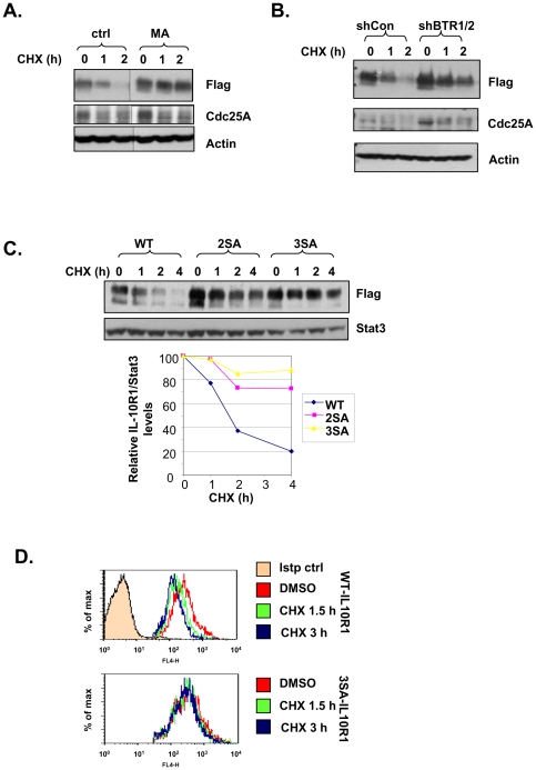
Figure 5. βTrCP-mediated ubiquitination drives IL-10R1 degradation.
(A) 293T cells stably transfected with IL-10R1 were pretreated for 0.5 h with or without 20 mM of methylamine (MA); (B) 293T cells stably expressing IL-10R1 were transfected with shCon or shBTR1/2; (C) 293T cells stably transfected with the WT, S319, 23A (2SA) or S319, 23, 70A (3SA) IL-10R1 were used; (D) 293T cells stably expressing WT or 3SA IL-10R1 were used. In (A), (B), (C) and (D), all cells were treated with 25 µg/ml of cycloheximide (CHX) for indicated times. In (A), (B) and (C), the levels IL-10R1-Flag in each sample were determined by immunoblotting. The levels of Cdc25A were used as an indicator of βTrCP knockdown efficiency (B). In (C), band intensities were determined by NIH ImageJ. Levels of IL-10R1 relative to those of Stat3 (loading control) were calculated and graphed (WT: diamonds; 2SA: squares; 3SA: triangles). In (D), cell surface IL-10R1 levels were determined by FACS analysis. Only cells stained positive for IL-10R1 were gated.

