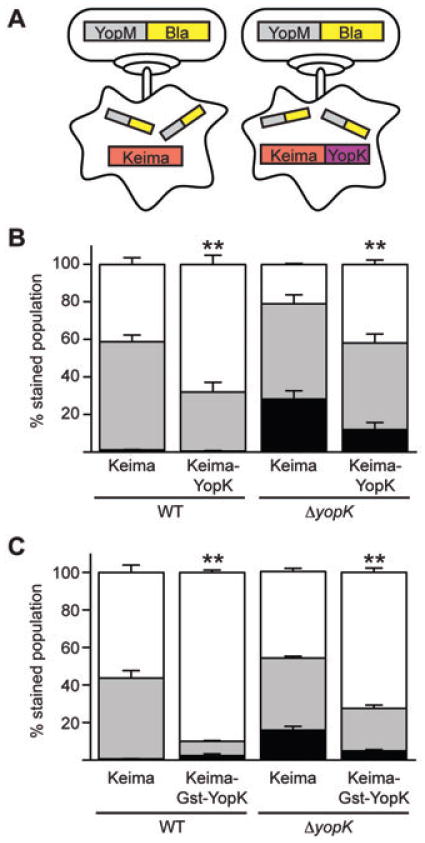
Fig. 6.

Keima–YopK negatively regulates effector Yop translocation. A. Schematic of transfection of CHO cells with plasmids expressing Keima or Keima–YopK followed by infection with Y. pestis and injection of the YopM-Bla reporter. B and C. CHO cells expressing either Keima, Keima–YopK or Keima–Gst–YopK were infected with WT or ΔyopK Y. pestis carrying the YopM-Bla reporter. The CHO cells were then stained with CCF2-AM and analysed by flow cytometry for red, green and blue fluorescence. Infections were performed in triplicate and are shown as averages with standard deviations. Student's t-test was performed to demonstrate significant differences in total injection levels (aqua + blue cells) for strains expressing Keima–YopK or Keima–Gst–YopK relative to the Keima-only controls (**P < 0.001). The data shown are representative of one data set, and the experiment was performed at least twice.
