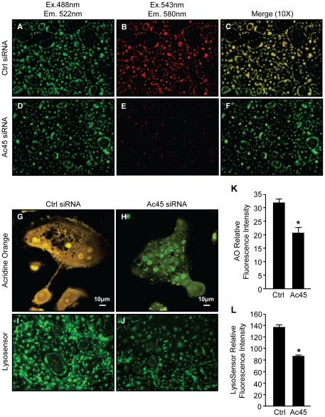
Figure 2. Ac45 is important for intracellular acidification in osteoclasts.
Acridine orange (AO) staining of RANKL-stimulated BMM-derived osteoclasts after Ctrl siRNA (A–C & G) or Ac45 siRNA (D–F & H) treatment for 48hrs under 10X magnification. Osteoclasts transfected with siRNAs were incubated with 5 µg/ml AO for 30mins at 37°C followed by confocal microscopy. Cells were first excited with wavelength of 488nm and emission wavelength of 522nm for unprotonated AO detection (A and D), followed by excitation wavelength of 543nm and emission wavelength of 580nm for protonated AO detection (B and E). Fluorescence shift of acridine orange from green to red indicate normal intracellular acidification. (C and F) Image merge of red and green fluorescence spectra of Ctrl and Ac45 siRNA treated cells. (G and H) Higher magnification (40X) of merged fluorescence spectra of a representative osteoclast from Ctrl and Ac45 siRNA treated population. (I and J) Osteoclasts were treated with 1 µg/ml LysosensorTM Green DND-189 for 30min and excited at wavelength of 488nm. (K and L) The fluorescence intensity was quantified for both acridine orange and LysosensorTM Green DND-189 and expressed as relative fluorescence intensity (n = 3). *P-values<0.05.

