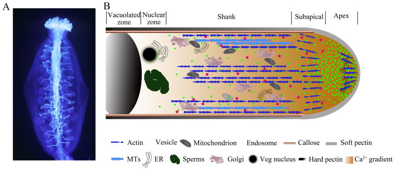
Figure 1. The pollen tube system: Directional and polarized cell growth.
(A) Pollen tubes in the pistil, aniline blue staining of pollen tubes in the Arabidopsis pistil. (B) Schematic diagram of the internal zonation and structural elements of a growing pollen tube. The growing tube displays a tip-focused cytoplamic Ca2+ gradient and contains a single soft pectin apical wall and two layers shank wall, the inner sheath of callose and outer coating of hard pectin, which are non-plastic and able to resist turgor pressure. The apical clear zone is characterized by a V shaped accumulation of secretory vesicles that facilitate massive tip-targeted exocytosis. The subapical organelle-rich zone is followed by a nuclear and a vacuole zone. Microtubules (MTs) and long actin cables axially aligned in the shank and are excluded from the apical zone. A collar-like actin microfilaments (F-actin) structure is present in the subapical region and a population of fine and short F-actin is detected in the extreme apex.

