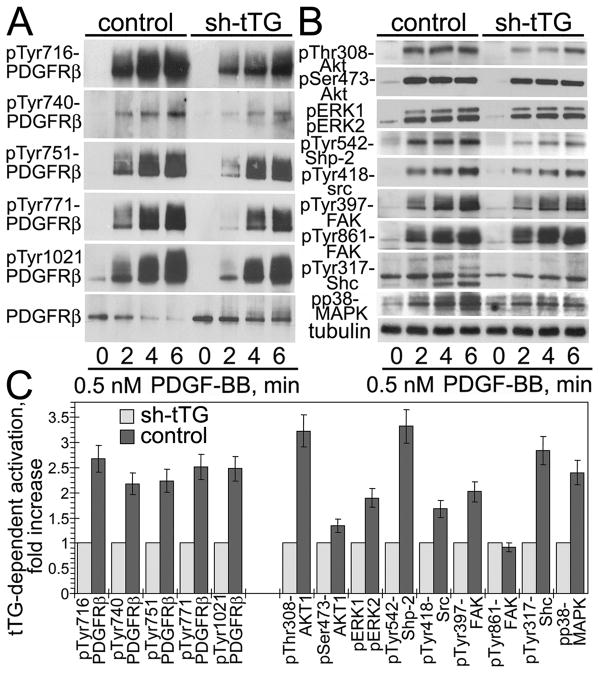
Figure 2. tTG increases PDGF-BB-dependent activation of PDGFRβ and its downstream signaling targets in vascular SMCs.
Adherent quiescent human aortic SMCs expressing non-silencing shRNA (control) or tTG shRNA (shtTG) were treated with 0.5 nM PDGF-BB for 0 6 min. (A,B) Activation levels of PDGFRβ (A) and its downstream targets (B) were defined by immunoblotting with antibodies to pTyr716-PDGFRβ, pTyr740-PDGFRβ, pTyr751-PDGFRβ, pTyr771-PDGFRβ, pTyr1021-PDGFRβ, PDGFRβ, pThr308-Akt1, pSer473-Akt1, pERK1/2, pTyr542-Shp-2, pTyr418-src, pTyr397-FAK, pTyr861-FAK, pTyr317-Shc, and pp38MAPK. All samples were normalized for equal amounts of tubulin. (C) Phospho-site signals were quantified, averaged, and expressed for control cells as -fold activation over those in shtTG-expressing cells. Shown are the means ± S.D. for three independent experiments.

