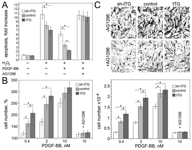
Figure 4. tTG stimulates PDGF-BB-induced survival, proliferation, and migration of vascular SMCs.
(A) tTG promotes PDGF-BB-mediated vascular SMC survival. Apoptosis of adherent quiescent human aortic SMCs expressing tTG shRNA (shtTG), non-silencing shRNA (control) or tTG was induced by treatment with 100 μM H2O2 for 12 hours with or without 10 nM PDGF-BB and 20 μg/ml AG1296. The numbers of apoptotic cells were quantified and expressed as -fold increase over those in the control cells without H2O2 and PDGF-BB. (B) tTG promotes PDGF-BB-mediated vascular SMC proliferation. Proliferation of adherent serum-starved human aortic SMCs expressing tTG shRNA (shtTG), non-silencing shRNA (control) or tTG was measured after induction with 0–10 nM PDGF-BB for 72 hours. Cell numbers were quantified and expressed as % change compared to those in the population of control cells without PDGF-BB. (C) tTG stimulates PDGF-BB-mediated migration of vascular SMCs. Chemotactic migration of 5×10⁴ 35S-labeled adherent quiescent human aortic SMCs expressing tTG shRNA (shtTG), non-silencing shRNA (control) or tTG through 8-μm membrane pores was studied in Transwells™ with 0–10 nM PDGF-BB in lower chambers. After 12 hours, transmigrated cells were removed from the filter bottoms; radioactivity was counted and converted to cell numbers. The upper panels show membrane undersides stained with crystal violet. Bar - 100 μm. (A–C) Some samples contained PDGFR inhibitor AG1296. Shown are the means ± S.D. for three independent experiments with measurements performed in triplicate, *P< 0.05.

