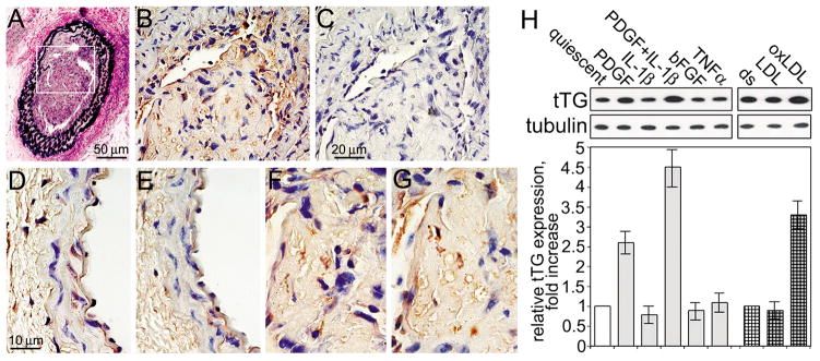
Figure 5. PDGF-BB and injury increase tTG levels in vascular SMCs.
(A–G) Left mouse carotid artery was wire-injured (Lindner et al., 1993) and neointima was allowed to form for 4 weeks. (A) Transverse section of the injury area was stained to visualize elastin. The area marked by rectangle shows neointima formation. (B,C) Extracellular tTG-generated Gln-Lys isopeptides are present in the neointima. Sections were labeled with mAb reacting with tTG-generated Gln-Lys isopeptide (B), or the same antibody preincubated with excess free Gln-Lys isopeptide (C). (D–G) The levels of tTG and extracellular tTG-generated cross-links are increased in neointima. Transverse serial sections of uninjured contralateral (D,E) and neointima in the injured (F,G) carotid arteries were labeled with antibodies to tTG (D,F) or tTG-generated Gln-Lys isopeptides (G,E). The staining was developed with peroxidase-conjugated secondary IgG, DAB substrate, and counter-staining with hematoxylin and eosin. Bars - 50 μm (A), 20 μm (B,C), and 10 μm (D–G). (H) PDGF-BB and oxidized LDLs increase tTG levels in human aortic SMCs. Quiescent adherent cells were treated with 2 nM PDGF-BB, 1 nM IL-1β, their combination, 2 nM basic FGF, or 1 nM TNFα for 48 hours (left panel). Adherent cells in delipidated 10% serum (ds) were treated with 100 μg/ml native or oxidized LDLs for 24 hours (right panel). The levels of tTG and tubulin were defined by immunoblotting. tTG amounts were quantified by densitometry, averaged, normalized for tubulin loadings and expressed as -fold increase over those in cells in quiescence (left panel) or in delipidated serum (right panel). Shown in (H) are the means ± S.D. for three independent experiments.

