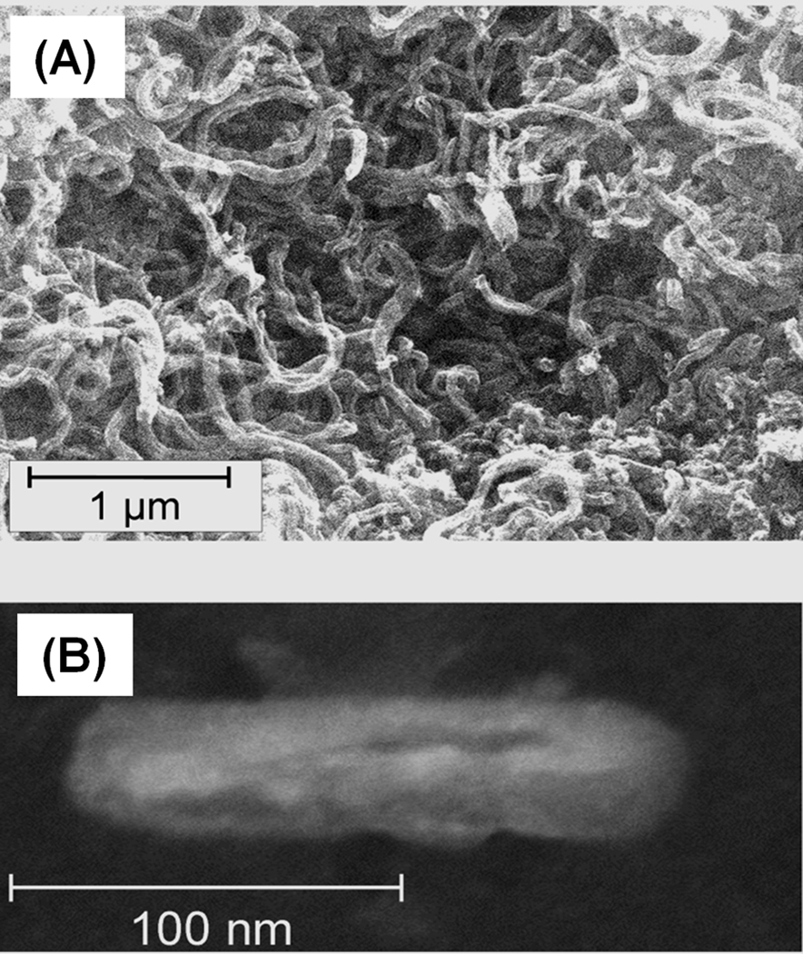
Fig. 18.
SEM image of pristine 1–2 µm long dry carbon nanotubes aggregated after exposure to water (A) and SEM micrograph of nanotubes oxidatively cut using treatment with a mixture of concentrated sulfuric and nitric acids (B). Adapted from ref. [94].

