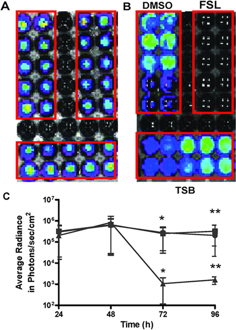
Fig. 5. Biofilm bioluminescence of Xen 43 decreased by farnesol.
Xen 43 biofilms were developed in 30 wells of an opaque 96-well microtiter plate (A). At 48 h, supernatants were removed and unadhered cells were washed with PBS. Three groups of ten wells each were exposed to DMSO, farnesol (FSL) (0.5 mM), or Trypticose soy broth (TSB) media for 24 h (B). Bioluminescence was monitored at 24, 48, 72 and 96 h and compared among the three exposures. Farnesol exposure significantly reduced average radiance compared with DMSO or TSB exposed wells (* and ** p < 0.05) (C). ■: DMSO; ●: TSB; ▲: Farnesol.

