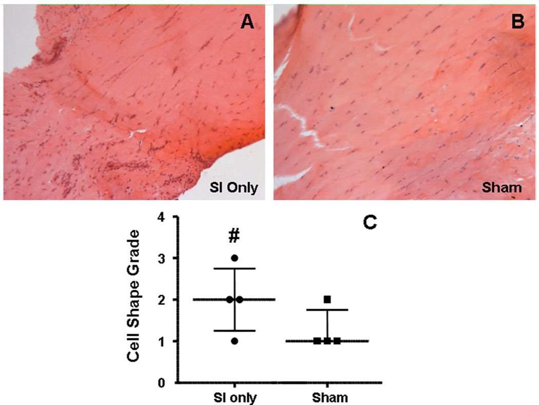
Figure 2.
After 1 week, a more rounded cell phenotype was seen in the intra-articular space with SI only (A) compared to sham (B). The median and inter-quartile ranges of the histological grading is seen in panel C. Also notice the more organized fibers in the sham image at this time point. (# denotes p<0.1)

