Figure 2.
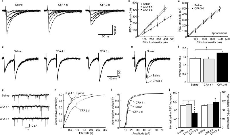
Persistent pain decreases GABAergic synaptic function by inhibiting presynaptic GABA release. (a) Representative traces of GABA inhibitory post-synaptic currents (IPSCs) evoked by various stimulation intensities in NRM neurons from a saline-injected rat and a CFA-injected rat at 4 h and 3 d post-injection. (b) A plot of input-output curves for IPSC amplitudes in neurons of the three treatment groups as in (a). Slopes: saline, 90.4 ± 11.5 pA 0.1 mA–1, n = 13; CFA 4 h, 88.7 ± 12.9 pA 0.1 mA–1, n = 26, p > 0.05; CFA 3 d, 53.6 ± 10.5 pA 0.1 mA–1, n = 35, p < 0.01. (c) A similar input-output plot of IPSCs from hippocampal CA1 neurons. Slopes: saline, 98.1 ± 14.8 pA 0.1mA–1, n = 15; CFA 3 d, 102.7 ± 16.2 pA 0.1mA–1, n = 16, p > 0.05. (d) Representative IPSC pairs evoked by two stimuli (100 ms apart) in NRM neurons from the three groups of rats. (e) Two representative IPSC pairs, from the two indicated groups, superimposed and scaled to the amplitude of the first IPSC. (f) Group data of changes in the paired-pulse ratios in the three groups (n = 15–35 cells for each group). (g) Representative traces of spontaneous miniature IPSCs (mIPSCs) in neurons from the three groups. (h–j) Distribution plots of mIPSC frequency (h) and amplitude (i) from neurons of each group and their group data (j, n = 16–20 cells).

