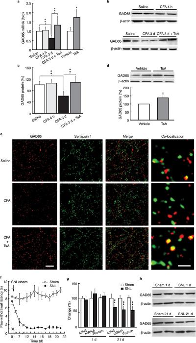
Figure 4.
Persistent inflammatory and neuropathic pain reduces GAD65 expression. (a) Effects of CFA and TsA on GAD65 mRNA levels in NRM tissues from control and CFA-injected rats (n = 5–7 each group). (b,c) Western lanes (b) and group data (c, n = 5– 7 each group) of GAD65 proteins in saline- and CFA-injected rats 4 h and 3 d post-injection without or with the TsA treatment in vivo. (d) Western lanes of GAD65 proteins in NRM tissues from normal rats. (e) Micrographs of immunohistochemical staining for GAD65 (red), the synaptic terminal protein synapsin I (green), their merged images and co-localization of GAD65 and synapsin I (yellow) in saline- (top row) and CFA- (bottom two rows) injected rats without or with the TsA treatment (n = 5–6 rats each group and 4–6 randomly selected sections were analyzed and compared). Scale bars = 50 μm (left three columns) and 2 μm (right column). (f) Time course of changes in pain threshold in rats after spinal nerve ligation (SNL, n = 6) or sham surgery (n = 5) performed on day 0 (arrow). (g) Normalized changes in levels of acetylated H3 at gad65 promoter (–646 to – 484 bp), GAD65 mRNA and protein in NRM tissues from sham and SNL rats at 1 d and 21 d after surgery (n = 5–6 each group). (h) Representative Western lanes of GAD65 and β-actin proteins from a sham rat and an SNL rat at 1 d and 21 d.

