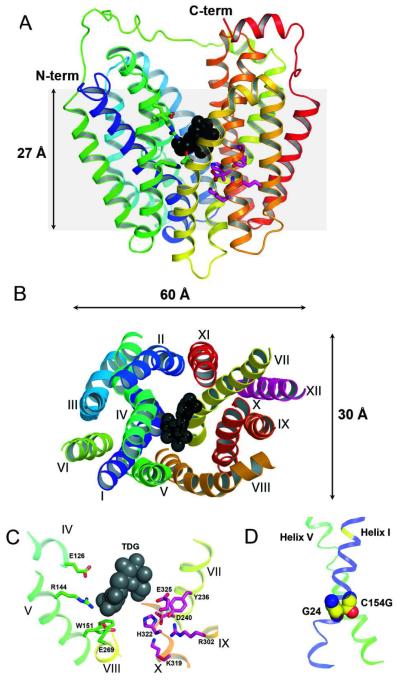
Figure 1.
X-ray structure of LacY with transmembrane helices rainbow colored from blue (helix I) to red (helix XII) and bound TDG presented as back spheres. Residues in sugar- and proton-binding sites are shown as green and pink sticks, respectively. (A) View parallel to the membrane (PDB ID 1PV7). Hydrophilic cavity is open to cytoplasm. Grey area represents the approximate thickness of the membrane phospholipid bilayer. (B) Cytoplasmic view showing dimensions of the LacY molecule and spatial packing of transmembrane helices. The loop regions are omitted for clarity. (C) Detailed view from cytoplasm showing residues in the sugar- and H+-binding sites. (D) Transmembrane helices I and V are tightly packed in the C154G mutant viewed parallel to the membrane (from LacY structure PDB ID 2CFQ). Gly residues 24 and 154 are shown as spheres.

