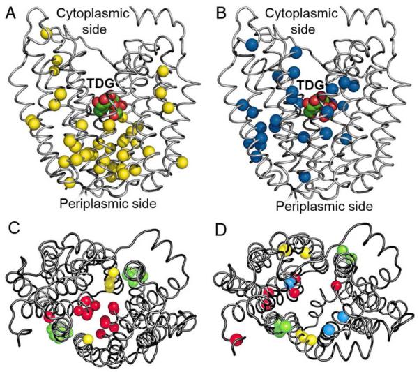
Figure 2.
Distribution of Cys replacements that exhibit changes in reactivity with NEM in the presence of TDG. Cα atoms of single Cys replacements are shown on backbone of the LacY structure in an inward-facing conformation (PDB ID 1PV7). (A and C) Positions of Cys residues that exhibit a significant increase in reactivity. (B and D) Positions of Cys residues that exhibit a significant decrease in reactivity. (A and B) Side view with bound TDG. (C and D) Cytoplasmic view demonstrating pseudo-symmetrical distribution of Cys replacements in putative translocation pathway. Residues located in symmetrically positioned helices are colored identically: I – VII (red); II – VIII (yellow); IV – X (blue); V – XI (green).

