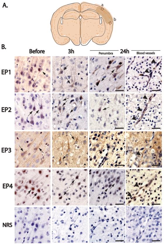
Figure 1. Immunohistochemistry of EP1-4 receptors in postnatal rat HIE brains.
(A) Schema of the cortical areas examined (box a: within infarct zone; box b: penumbra). EP1-4 receptor expression was examined in cerebral cortex in layers II/III dorsally and ventrally to the infarct area in the penumbra at 3h and 24h after HI. (B) EP1-4 receptors are dynamically regulated in sham (just before onset of hypoxia) and HI (3h and 24h after hypoxia) pups in cerebral cortical neurons (thin arrows) and microvasculature (arrowheads). Normal rabbit serum (NRS) control stains did not show specific staining. Scale bar= 50μm

