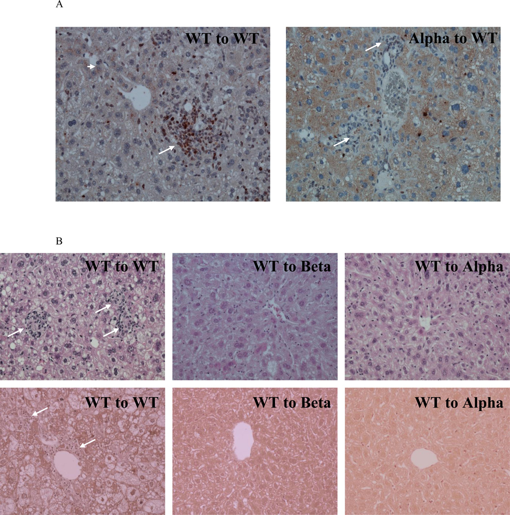
Figure 6. Expression of IL-1 in bone marrow-derived cells did not promote steatohepatitis and liver fibrosis development in IL-1α- or IL-1β-deficient mice.
Liver specimens were obtained from the specified experimental groups as described in figure 5. (A) Representative images of immunohistochemistry staining for IL-1α in livers from WT to WT and Alpha to WT mice. WT to WT livers showed increased staining of IL-1α protein both in hepatocytes (Arrow head) and in cells within inflammatory foci (Arrows). Livers from Alpha to WT also showed increased expression in hepatocytes (Arrow head) but showed a substantially lower number of cells with positive IL-1α staining in inflammatory foci (Arrows). (B) Representative images of H&E (upper panel) and Masson trichrome (lower panel) staining of livers from WT to WT, WT to Beta, and WT to Alpha mice are shown. Arrows in H&E staining show inflammatory foci. Arrows in Masson staining show collagen deposition. Magnification ×200.

