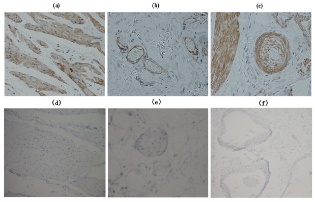
Figure 2.
Immunohistochemical analysis of EF-Tu expression in interstitial tissue: (a) Smooth muscle; (b) venous endothelial cells; (c) artery endothelial cells, showed high EF-Tu expression. Immunohistochemical analysis of EF-Tu negative control: (d) Smooth muscle; (e) venous endothelial cells; (f) artery endothelial cells.

