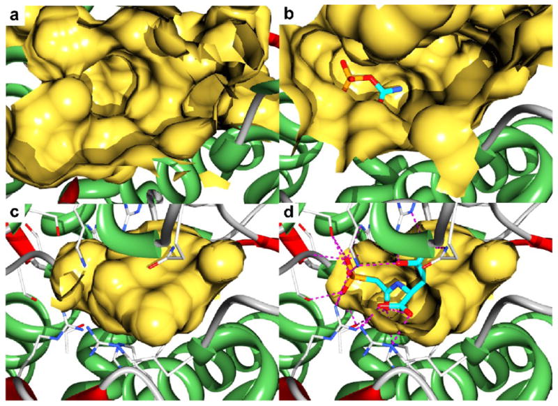
Fig. 3.

X-ray structures of the B. subtilis ATCase showing the active site cavity as calculated by Castp15 (a-d). (a) The active site of the enzyme in the absence of ligands. (b) The active site of the ATCase•CP complex with CP shown as sticks. (c) The active site of the ATCase•PALA complex. (d) A cut away view into the active site cavity of the ATCase•PALA complex showing the interactions (dotted lines) between the active site residues and PALA (sticks).
