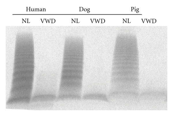
Figure 1.

Multimer distribution of von Willebrand factor antigen in normal (NL) and von Willebrand disease (VWD) human, dog, and pig plasma samples. Human plasmas were obtained from George King Bio-Medical, Inc., Overland Park, Kansas. The human VWD plasma was from a patient with severe type 3 VWD plasma with VWF : RCo 15 IU/dl and VWF : Ag 1 IU/dl. The dog and pig plasmas were prepared at the Francis Owen Blood Research Laboratory at the University of North Carolina at Chapel Hill using normal and severe VWD animals that had no detectible activity or antigen in either species. None of the subjects had recently been transfused with VWF-containing products. Anti-VWF antibodies for immunostaining were purchased from Dako (A082, Carpintera, CA) (1.5% agarose gel) [7–9]. The identity of the very bottom band seen in all lanes is unknown but is a consistent finding in multiple laboratories and is also seen in murine VWD plasma [7–11]. It is possible that this band simply represents nonspecific binding of the antibody to the leading edge of the proteins at the end of the electrophoresis procedure.
