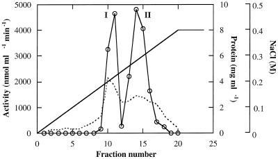
Figure 2.
Separation of alkaline α-galactosidase forms I and II on Mono-Q chromatography. The fractions active at pH 7.5, represented in Figure 1, were pooled, desalted, applied to the column, and eluted with the indicated linear gradient of 0.1 to 0.4 m NaCl. Activity of each fraction was assayed with pNPG at pH 5.0 and pH 7.5.

