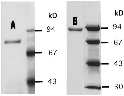
Figure 8.
The purified alkaline α-galactosidase form I (lane A) and II (lane B) in SDS-PAGE gel showing the denatured molecular masses of 79 kD and 92 kD, respectively. The partially purified proteins, described in Table II, were further purified with steps of native electrophoresis, as described in Methods. Next to each of the purified proteins is a lane showing the separation of markers of known molecular mass.

