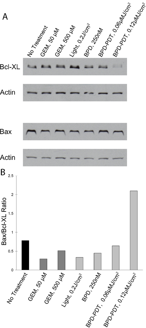
Figure 5.
Western blots for Bcl-XL and Bax protein content. Cells were treated with sub-curative doses of PDT and gemcitabine, and extracts were probed for Bcl-XL and Bax protein content by Western blot. In (A), from left to right, the lanes in the blot correspond to untreated cells, 50 µM gemcitabine, 500 µM gemcitabine, 0.2 J/cm2 light dose only (no verteporfin), 250 nM verteporfin only, no light, verteporfin PDT with 0.1 J/cm2 light dose, and with 0.2 J/cm2 light dose. In (B), the ratio of Bax to Bcl-XL is shown from densitometry analysis of the western blot data in (A).

