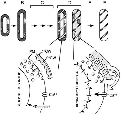
Figure 10.
Model of the coordination of PCD and secondary cell wall synthesis during TE differentiation. A, Mechanically isolated mesophyll cells were induced to differentiate with auxin and cytokinin. B, After 24 h, the cells expand and some cells divide (not a prerequisite for differentiation). C, A number of molecular events presumably precede visible manifestations of differentiation, including the synthesis of hydrolytic enzymes that are likely sequestered in the vacuole. D, At approximately 72 h, differentiation is visibly manifested by the appearance of secondary cell wall thickenings. A 40-kD Ser protease (represented by dots in vesicles and cell wall) is secreted concomitantly with secondary cell wall materials (shaded vesicles), leading to an increase of extracellular protease activity as secondary cell wall synthesis proceeds. Approximately 6 h after the first appearance of secondary cell wall thickenings, a critical activity of protease is reached in the extracellular matrix, which triggers cell death, ending secondary cell wall synthesis. Cell death is initiated by an influx of Ca2+, leading to vacuole collapse and cessation of cytoplasmic streaming. E, Mixing of the hydrolytic vacuole with the cytoplasm leads to autolysis. F, The cell is completely cleared within 8 to 12 h, leaving the functional cell corpse composed of secondary cell wall. The secondary cell wall, represented here as having a stylized reticulate pattern, is drawn separated from the plasma membrane for clarity. 1°CW, Primary cell wall; 2°CW, secondary cell wall; PM, plasma membrane.

