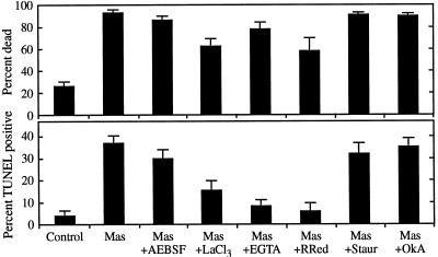
Figure 5.
Inhibition of mastoparan-induced cell death and DNA fragmentation by antagonists of Ca2+ influx. Five-hundred-microliter aliquots of cultures containing nascent TEs were pretreated for 30 min with 0.1 mm 4-(2-aminoethyl)-benzenesulfonyl fluoride (AEBSF; a Ser protease inhibitor), 150 μm LaCl3 (a Ca2+ channel antagonist), 500 μm ethylene glycol-bis(β-aminoethyl ether)-EGTA (a Ca2+ chelator), 50 μm ruthenium red (RRed; a Ca2+ channel antagonist), 10 μm staurosporine (Staur; a protein kinase inhibitor), or 40 nm okadaic acid (OkA; a protein phosphatase inhibitor), and were then treated with 2.5 μm mastoparan (Mas). The percentage of dead cells was determined 1 h later using fluorescein diacetate; the percentage of cells exhibiting DNA fragmentation was determined using TUNEL 3.5 h after treatment. Control cells were not treated with drugs before processing. Error bars represent the se of two samples treated in parallel.

