Figure 12.
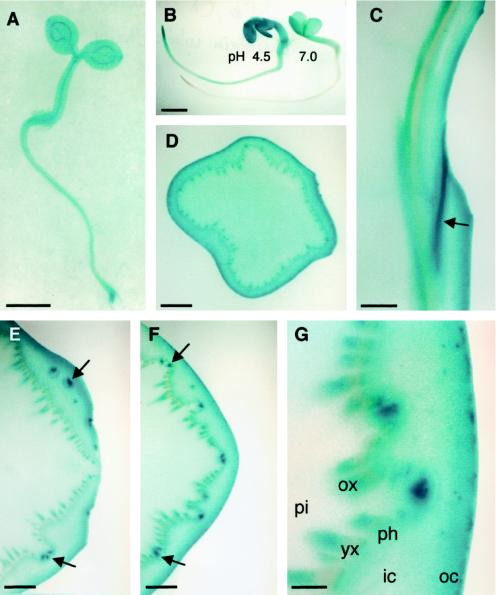
In situ activity of α-glucosidase assayed with α-X-gluc at pH 4.5 for 12 h, except where noted. A, Three-day-old Arabidopsis seedling. B, Three-day-old mustard seedlings stained at pH 4.5 (left) or 7.0 (right) for 3 h. C, Longitudinal section through a broccoli stem. The curved surface on the right side is a leaf abscission zone. Unstained tissue on the left is pith. Strong staining is seen in the leaf trace indicated by the arrow. D, Cross-section of a broccoli stem approximately 3 cm from the apex. E, Cross-section of a broccoli stem through a leaf abscission zone. Arrows point to strong staining in leaf traces at or above the point where they separate from the vascular cylinder. A leaf abscission zone is on the right side of the image. F, Same as E but approximately 4 mm basal. Arrows point to the same leaf traces as in E, but both leaf traces are below the point of separation from the vascular cylinder. G, Finer detail of F. pi, Pith; ox, older xylem; yx, young xylem; ph, phloem; ic, inner cortex; oc, outer cortex. Note the increased staining toward the outer cortex and in the older xylem and in the leaf traces. Bars = 1 mm (A, B, and D); 2 mm (C, E, and F); or 0.5 mm G.

