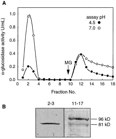
Figure 5.
Separation of α-glucosidases from an Arabidopsis stem+bud extract by ConA chromatography and an immunoblot of the pooled active fractions probed with anti-Aglu-1. A, ConA-Sepharose column. Bound proteins were eluted with 15 mm methyl Glc (MG). α-Glucosidase activity was measured at pH 4.5 (•) and pH 7.0 (○). U, Unit. B, Immunoblot of activity peaks separated in A. Nonbinding activity (fractions 2 and 3) and bound activity (fractions 11–17) from A was concentrated 24-, and 215-fold, respectively, and equal volumes of each concentrated fraction were separated by SDS-PAGE and probed with anti-Aglu-1.

