
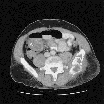
FIG. 2. Axial contrast-enhanced CT scan of the abdomen showing intussusception (A) in the right lower quadrant. A large mixed lytic and sclerotic lesion in the left hemi-pelvis (B) extending to the sacroiliac joint, typical of multiple myeloma, was noted.