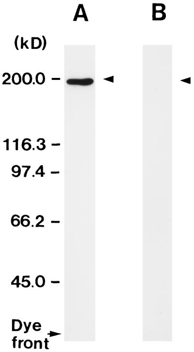
Figure 1.
Western blot showing the cross-reactivity of single-affinity-purified anti-NADH-GOGAT IgG to NADH-GOGAT protein of rice roots. A, Immunoblot of a crude extract from rice roots (10 μg) labeled with single-affinity-purified anti-NADH-GOGAT IgG. B, Same as A except that the NADH-GOGAT IgG was preabsorbed with an excess amount of the NADH-GOGAT protein purified from cultured rice cells prior to immunolabeling. The position and sizes (in kD) of protein markers are indicated at the left.

