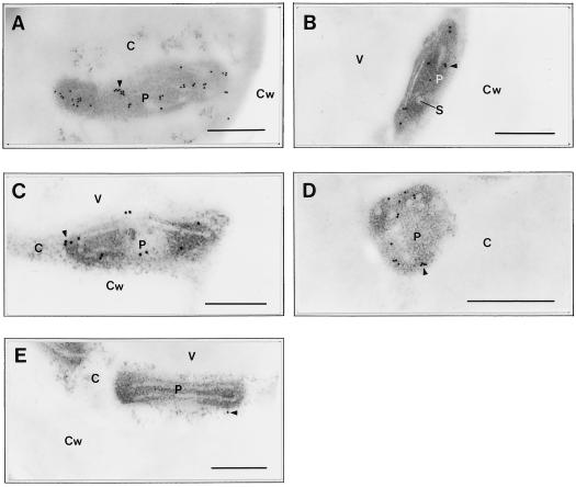
Figure 4.
Immunolabeling of NADH-GOGAT protein in different populations of plastids in rice roots. Sections were labeled with single-affinity-purified anti-NADH-GOGAT IgG using tissue from a seedling grown with 1 mm NH4+ for 24 h and sampled from the tip (<10 mm). A and B, Epidermis; C through E, vascular parenchyma. Note the different ultrastructures of the five representative plastids shown: no defined internal structures (A), a well-defined starch grain and internal lamellae (B), a single internal lamella (C), globular, membranous structures (D), and well-developed internal lamellae (E). Sections shown in A through D were incubated with single-affinity-purified anti-NADH-GOGAT IgG as the primary antibody. The section shown in E (the control) was incubated with anti-NADH-GOGAT IgG pretreated with an excess amount of NADH-GOGAT protein. P, Plastid; C, cytosol; Cw, cell wall; S, starch grain; V, vacuole. Arrowheads indicate gold label. Bars = 0.5 μm.

