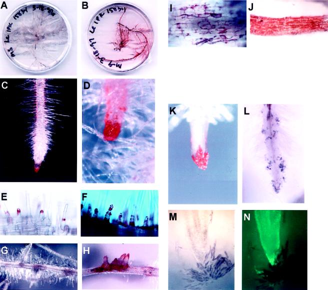
Figure 2.
L. erythrorhizon hairy-root cultures showing pigment formation in different cells under varying conditions. A, Transformed roots growing on M medium. B, Transformed roots growing on M-9 medium. Pigment diffusion from the roots into the medium was apparent at 3 weeks. C, Root tip grown on M medium showing normal pigment production pattern in border cells and root hairs. D, Root tip grown on M-9 medium showing increased pigmentation in border cells and root hairs. E, Pigment deposition patterns in M-grown roots. Emerging root-hair tips have a cap of pigment, which remains at about that same distance from the epidermis as the root hair grows. F, Root hairs of M-9-grown roots showing exudation of pigment in droplets all over the hairs. G, Lateral root formation in M-grown roots. Pigment is apparent in the region of root emergence. H, Lateral root formation in M-9-grown roots. Numerous roots emerge at a single point and pigmentation is more intense than in M-grown roots. I, Pigment deposition in a few of the cells near lateral root eruption. J, Pigment production in all epidermal cells of M-9-grown root. K, Border cell pigmentation on root cap of M-grown root. Pigments are confined to border cells. L, Border cell deposition along growing root in solid M medium. M, Light micrograph of normal (untransformed) root tip placed in water showing dispersion of border cells from the cap. Border cells are purple, cap is white. N, UV fluorescence of root stained with fluorescein diacetate showing living cells of root. Most border cells do not take up the stain.

