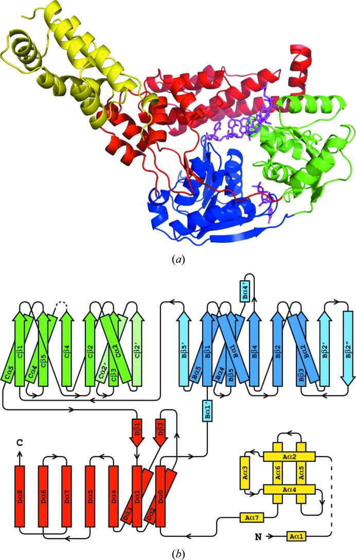
Figure 1.
(a) The structure of hSuv3 with its four domains colored as follows: the N-terminal domain A is shown in yellow, the two RecA domains B and C are shown in blue and green, respectively, and the C-terminal domain D is shown in red. Both AMPPNP and RNA are positioned as in their individual complexes and shown in magenta. (b) Topology of hSuv3. The secondary-structure elements of domains B and C which are not present in the canonical topology of RecA are represented in pale colors.

