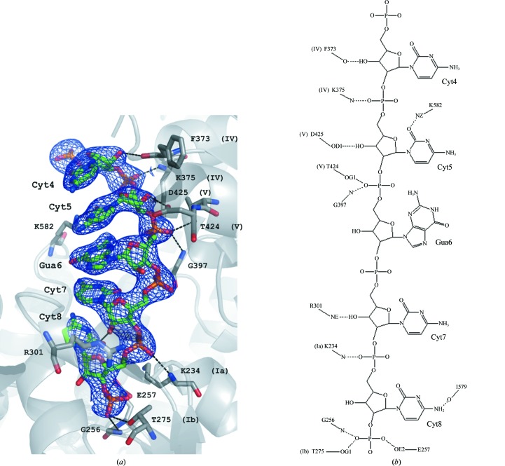
Figure 3.
(a) The identified fragment of nucleic acid in the RNA–hSuv3 structure in F obs electron density (at a 1.4σ contour level). The surrounding protein is shown in cartoon representation, with residues that interact with RNA shown in ball-and-stick representation. (b) The scheme of hydrogen bonds identified between the RNA oligonucleotide and hSuv3. Roman numerals refer to the helicase motifs of particular residues.

