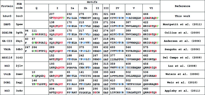
Figure 4.
Structurally aligned sequences of typical helicase motifs in hSuv3 and selected SF2 family helicases from the PDB containing both ATP analogs and fragments of nucleic acids. Residues hydrogen bonded to ATP are shown in red, those hydrogen bonded to nucleic acid are shown in blue and those stacked with adenine are shown in green. Residues in a left-handed α-helical conformation are underlined.

