Figure 4.
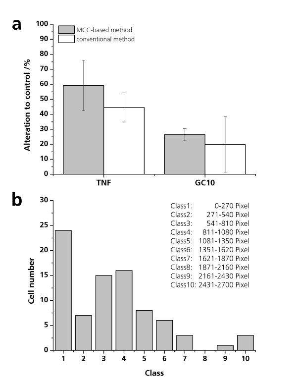
GFP expression of TNF-alpha- and GC10-exposed pIL8-GFP A549 cells. pIL8-GFP A549 cells were cultured in the microcavities and exposed to 20 ng/ml TNF-alpha or 30 μg/ml GC10 for 24 h under physiological conditions. In parallel, 10,000 pIL8-GFP A549 cells were seeded in 96-well microplates and stimulated with 20 ng/ml TNF-alpha and 30 μg/ml GC10 for 24 h. After the exposure time, the GFP expression of the pIL8-GFP A549 cells in the microcavities was analyzed by fluorescence microscopy. The percentage of GFP-expressing cells in the microcavity was calculated via the software analysis. The GFP expression of the pIL8-GFP A549 cells in the 96-well micro plate was quantified by fluorescence spectrometry. The percentage of GFP expression is pictured as alteration to the untreated control (alteration to control/percent). (b) pIL8-GFP A549 cells were treated for 24 h with 20 ng/ml TNF-alpha in the microcavities. The GFP expression of 90 individual cells was quantified. The classes of the fluorescence intensities (x-axis: class of GFP intensity) and its frequency (y-axis: frequency) is presented.

