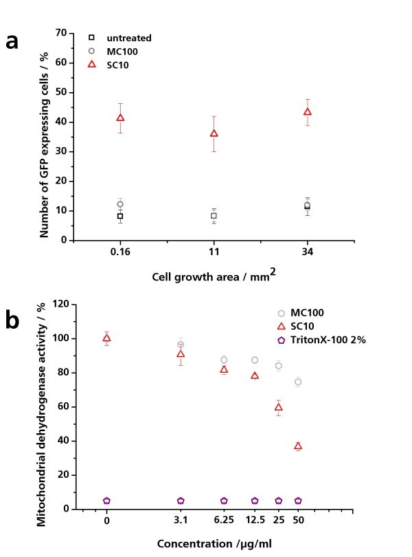
Figure 6.
Effect of nanoparticles on pIL8-GFP A549 cells. (a) Effect of miniaturization on nanoparticle-induced inflammation in pIL8-GFP A549 cells. pIL8-GFP A549 cells were cultured on three different growth areas (0.16, 11, and 34 mm2) and exposed to 20 μM SC10 and 20 μM MC100 for 24 h under physiological conditions. The percentage of GFP-expressing cells per growth area was analyzed by fluorescence microscopy. The amount of GFP-expressing pIL8-GFP A549 cells in relation to the cell growth area is depicted. The results are presented as mean of three independent experiments ± SD. (b) Concentration-dependent effect of nanoparticles on mitochondrial dehydrogenase activity of pIL8-GFP A549 cells. pIL8-GFP A549 cells were exposed to 0 to 50 μg/ml SC10 und MC100 for 24 h under physiological conditions. Triton X-100 was used as positive control. Via WST-1 assay the mitochondrial dehydrogenase activity was quantified. Untreated cells were set as 100%. The results are presented as mean of three independent experiments ± SD compared to the untreated control.

