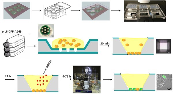
Figure 8.
Schematic experimental design. To reduce the evaporation of the cell culture medium, a biocompatible cell culture chamber was positioned on the top of the MCC. Each chamber of the covered silicone FlexiPerm® chamber includes seven individual miniaturized microcavities for statistical analysis of the experimental data. For each experiment, 100 μl cell suspension (100,000 cells/ml) were placed in each of the six culture segments. After 30 min, the cells adhered on the Si3N4 membrane. The segments of the cell culture chamber were filled with 100 μl cell culture medium. After 24 h of cell proliferation, the cells were exposed to the nanoparticles by aspirating the medium, washing the cells with PBS, and adding the nanoparticle-containing medium. After the exposure time, the cells were analyzed microscopically.

