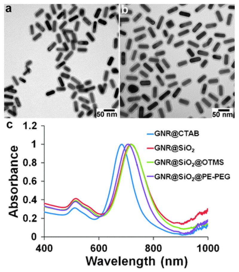
Fig. 2.

TEM and spectroscopic characterization of caged GNRs. (a and b) TEM images of GNR@SiO2 and GNR@SiO2@PE-PEG. The silica shell is 7.5 nm, and the organic molecules on the silica shell surface are not visible because they are not electron-dense materials. (c) Spectroscopic monitoring of GNR encapsulation. Initial CTAB capped GNRs dispersed in water show two extinction peaks at 515 (transverse) and 685 nm (longitudinal). Subsequent surface modifications with an SiO2 shell (dispersed in ethanol), C18 (dispersed in CHCl3) and PE-PEG (dispersed in water) shift the longitudinal peak to 719, 720, and 708 nm, respectively.
