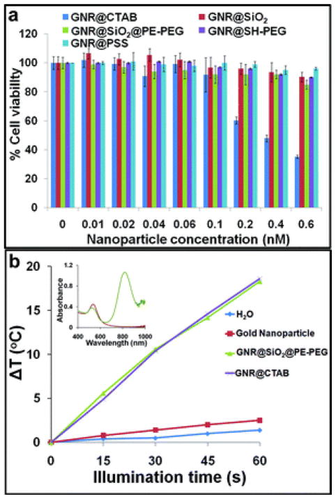
Fig. 5.
Characterization of cytotoxicity and capability of photothermal conversion of GNR@SiO2@PE-PEG. (a) Dose-dependent cytotoxicity of GNR@SiO2@PE-PEG in comparison with GNR@CTAB, GNR@PSS, GNR@SH–PEG, GNR@SiO2 in LNCaP prostate tumor cells. In the concentrations probed (0–0.6 nM), GNR@CTAB show high toxicity but not the other samples (24 h incubation of cells with the various GNRs). (b) Rates of temperature increase (ΔT) for GNR@CTAB, GNR@SiO2@PE-PEG, spherical gold nanoparticles, and water during NIR-laser illumination. When fitted with linear trendlines, GNR@CTAB and GNR@SiO2@PE-PEG show nearly identical performance and their temperature increase rates are approximately 7 and 13 times faster than those of gold nanoparticles and water. (Inset) Vis-NIR extinction spectra of the GNR@SiO2@PE-PEG and spherical gold nanoparticles of 18 nm.

