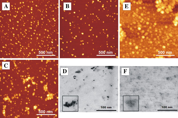
Figure 4.
AFM (a, b, c, e) and STEM (d, f) images of CdTe quantum dots. a–c AFM images of quantum dots dispersed on mica (dispersed from aqueous solution kept for: a 40 min, b 5 h, c 24 h), d STEM image of quantum dots dispersed on TEM grid (dispersed from solution kept for 48 h), e AFM image of quantum dots with BSA dispersed on mica (dispersed from aqueous solution kept for 2 months), f STEM image of quantum dots with BSA (dispersed from aqueous solution kept for 2 months). Inserts (in d and f images) show magnified view (40 nm × 40 nm). Concentrations of solutions used for sample preparation were 6 × 10⁻⁶ mol/l.

