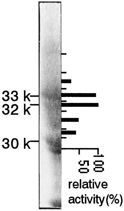
Figure 4.
Preparative SDS-PAGE was carried out using 11% acrylamide slab gels. A portion of the gel in this figure was stained with Coomassie brilliant blue and the rest of the gel was stained with Cu. The gel containing proteins between 30 and 35 kD in size was cut into seven fragments (indicated by the short lines). The recovery was low but NAS activity was reproducibly detected by elimination of SDS and Cu during electroelution of peptide from the gels. The thick bars indicate relative NAS activity of peptides from each gel fragment. The activity of the most active fraction was set at 100%.

