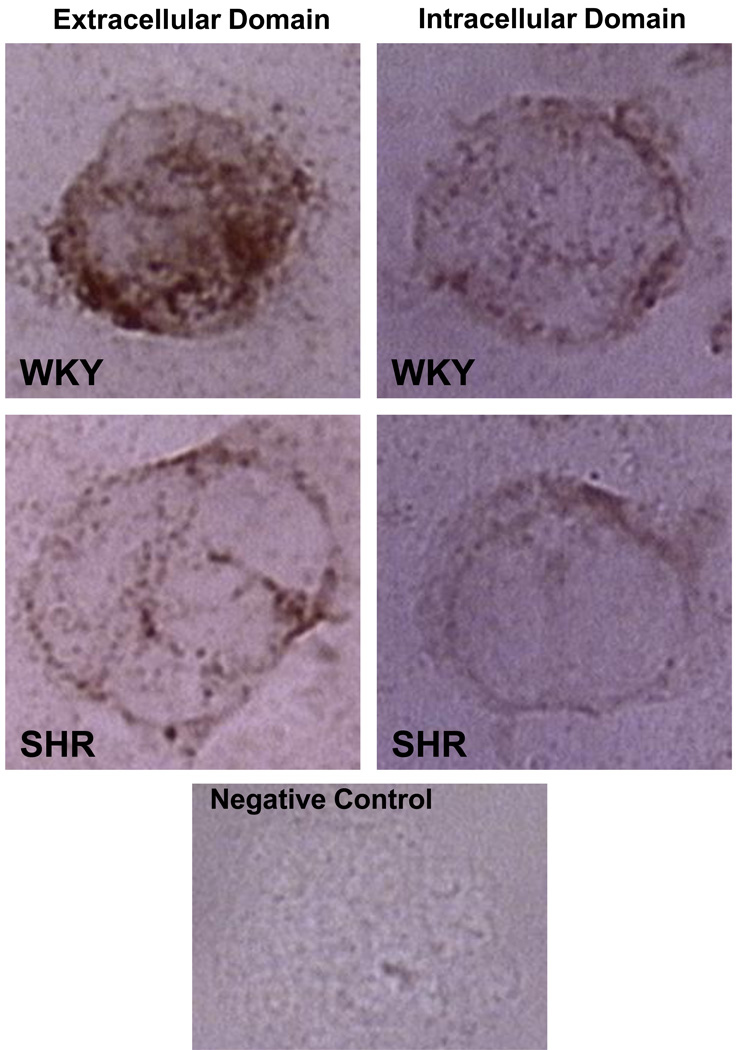
Figure 3.
Selected micrographs of the formyl peptide receptor (FPR) density on typical WKY and SHR leukocytes after immunolabeling with an antibody against its extracellular (left images) and intracellular domain (right images). The images are representative for the average receptor density as measured previously (74). Bottom image shows control label density without primary antibody. Note the reduced extracellular labeling density on SHR leukocytes as compared to the normotensive WKY, a trend that is not present when an antibody against the intracellular domain is used, suggesting FPR cleavage in the SHR.

