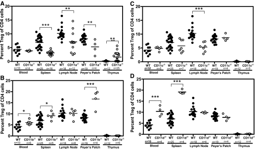
Figure 3.
Treg frequency in lymphoid tissues in wild-type and β2-integrin-deficient mice. Blood, spleen, lymph nodes, Peyer’s patches, and thymus were harvested from C57BL/6 mice; cells were isolated using a Miltenyi Treg kit, as described previously [49]; and Tregs (CD4+CD25+FoxP3+) were analyzed using flow cytometry. Data shown are gated on CD4+ cells. Statistical analysis was performed for each wild-type and β2-integrin-deficient tissue using the Student’s t-test (**, P≤0.01; ***, P≤0.001). (A) CD11a−/−mice. (B) CD11b−/−mice. (C) CD11c−/−mice. (D) CD11d−/−mice.

