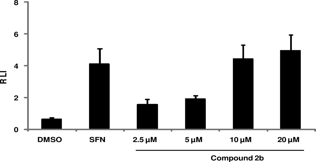
Figure 3. Levels of NQO1-ARE luciferase activity after treatment with compound 2b.
NQO1-ARE luciferase activity was measured by using stably transfected Beas-2B cells after treatment with compound 2b or sulforaphane (SFN) or dimethyl sulfoxide (DMSO). The exposure to compound 2b resulted in a significant concentration-dependent increase in luciferase activity as relative luminescence intensity (RLI). Data are representative of 3 independent experiments. Values shown are mean ± SD of quadruplicate wells (P ≤ 0.05).

